View All Electron Microscopy at Instruct
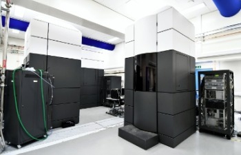
The Electron Bio-Imaging Centre (eBIC) provides scientists with state-of-the-art experimental equipment and expertise in the field of cryo-electron microscopy, for both single particle analysis and cryo-tomography. Currently eBIC houses five Titan Krios microscopes, a Talos Arctica, Glacios, and a Scios and Aquilos cryo-FIB/SEM.
Titan Krios I, II, III, IV & V are state of the art 300 kV cryo-electron microcopes, equipped with the latest generation of direct electron detectors. Krios I & II are equiped with the FEI Falcon III and the Gatan Bio-quantum K2 summit. Krios III, IV and V are equipped with the Gatan Bio-quantum K3 summit. The Falcon III and K2 detectors are intergated into FEI's automated data aquisition softwares for single particle and tomography, EPU and TOMO4 respectively. Krios I to IV can also be operated using SerialEM. The Talos Arctica is a new generation fully automated 200 kV FEG electron microscope. It is equipped with an autoloader system that permits loading of up to twelve specimens, automated data collection at liquid nitrogen temperature, and features a Constant-PowerTM objective lens for optimal contrast/resolution balance. The Scios is a state-of-the-art DualBeam™ Scanning Electron Microscope (SEM) and Focused Ion Beam (FIB) system that allows precision milling of samples. Technological advances have increased throughput and milling precision, yielding higher quality results in less time, making it an ideal system for thinning of samples for subsequent TEM imaging. The Thermo Scientific™ Aquilos™ Cryo-FIB is the first cryo-DualBeam™ (focused ion beam/scanning electron microscope) system dedicated to preparation of frozen, thin lamella samples from biological specimens for high-resolution tomographic imaging in a cryo-transmission electron microscope (cryo-TEM). Cryo-electron tomography’s ability to visualize structures in their native context allows researchers to observe functional relationships and interactions with other components in the cellular environment. This technique promises to become an important tool for scientists seeking a better understanding of living systems at the molecular level.
Harwell Science & Innovation Campus
OX11 0DE, Didcot
United Kingdom